
File:Mitosis-fluorescent.jpg
| |
This is a file from the Wikimedia Commons. Information from its description page there is shown below.
Commons is a freely licensed media file repository. You can help. |
Summary
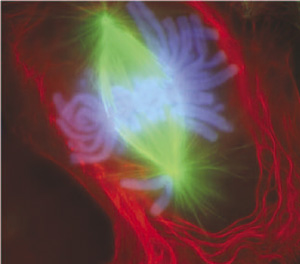
An image of a newt lung cell stained with fluorescent dyes undergoing mitosis, specifically during early anaphase. According to NIH, "The scientists use newt lung cells in their studies because these cells are large, easy to see into, and are biochemically similar to human lung cells."
The material stained green are the mitotic spindles, the material stained red is the cell membrane and some components of the cytoplasm near it, and the material stained light blue are the chromosomes.
From http://publications.nigms.nih.gov/moleculestomeds/images/newtcells.jpg - so it's public domain as it's US government material.
Licensing
 |
This image is a work of the National Institutes of Health, part of the United States Department of Health and Human Services. As a work of the U.S. federal government, the image is in the public domain. |
 |
| Annotations | This image is annotated: View the annotations at Commons |
File usage
I want to learn more...
SOS Children's Villages chose the best bits of Wikipedia to help you learn. By supporting vulnerable children right through to adulthood, SOS Children's Villages makes a lasting difference to the lives of thousands of people. Education is a key part of our work, and our schools provide high-quality teaching to the children in our care. There are many ways to help with SOS Children's Villages.