 |
This is a file from the Wikimedia Commons. Information from its description page there is shown below.
Commons is a freely licensed media file repository. You can help.
|
| Description |
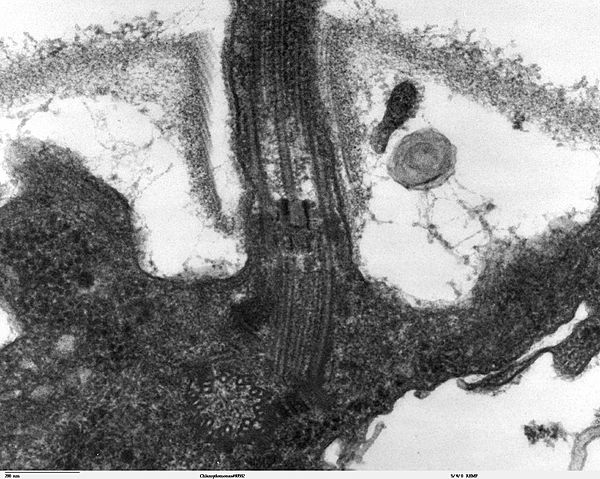
Transmission electron microscope image, showing an example of green algae (Chlorophyta).
Chlamydomanas reinhardtii is a unicellular flagellate used as a model system in molecular genetics work and flagellar motility studies.
This image is a longitudinal section through the flagella area. In the cell apex is the basal body that is the anchoring site for a flagella. Basal bodies originate from and have a substructure similar to that of centrioles, with nine peripheral microtubule triplets(see structure at bottom centre of image). The two inner microtubules of each triplet in a basal body become the two outer doublets in the flagella. This image also shows the transition region, with its fibers of the stellate structure. The top of the image shows the flagella passing through the cell wall. |
| Date |
20 September 2007 |
| Source |
Source and public domain notice at http://remf.dartmouth.edu/imagesindex.html |
| Author |
Dartmouth Electron Microscope Facility, Dartmouth College |
Permission
( Reusing this file) |
Released into the public domain
|
| Public domainPublic domainfalsefalse |
 |
This work has been released into the public domain by its author, Dartmouth Electron Microscope Facility, Dartmouth College. This applies worldwide.
In some countries this may not be legally possible; if so:
Dartmouth Electron Microscope Facility, Dartmouth College grants anyone the right to use this work for any purpose, without any conditions, unless such conditions are required by law.Public domainPublic domainfalsefalse
|
File usage
The following pages on Schools Wikipedia link to this image (list may be incomplete):
Through Schools Wikipedia, SOS Children's Villages has brought learning to children around the world. SOS Children cares for children who have lost their parents. Our Children's Villages give these children a new home and a new family, while a high-quality education and the best of medical care ensures they will grow up with all they need to succeed in adult life. Why not try to learn more about child sponsorship?


